
Lightning Flash 3.0
The Next Evolution in Lightning Flash Technology
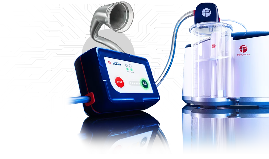
Part of Penumbra’s CAVT™ Platform (Computer Assisted Vacuum Thrombectomy)
Lightning Flash 3.0
Designed for the rapid removal of pulmonary and venous thrombus
- Faster clot removal timea
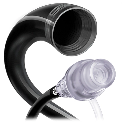
- Designed for large thrombus with enlarged tubing
- Automatic decompression engineered for uninterrupted clot removal
- Increased algorithmic sensitivity to thrombus and blood
- Engineered to reduce EBL
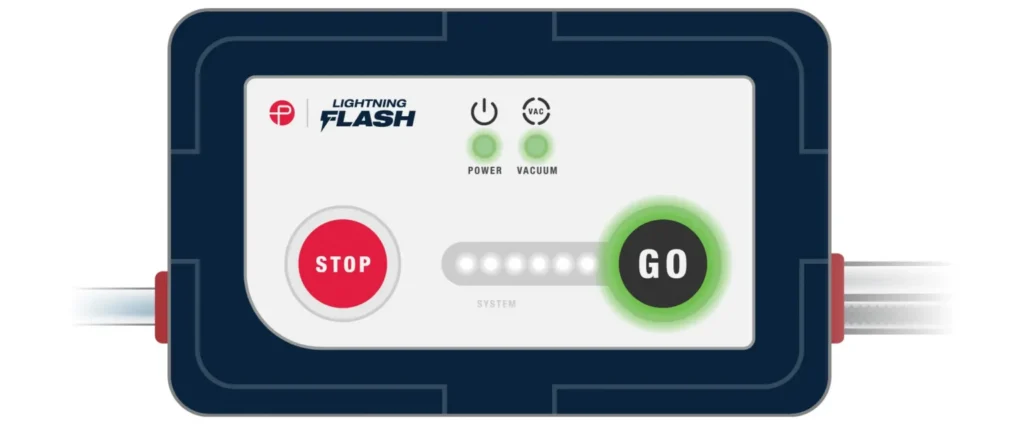
- Upgraded user interface
- Streamlined audio-visual feedback
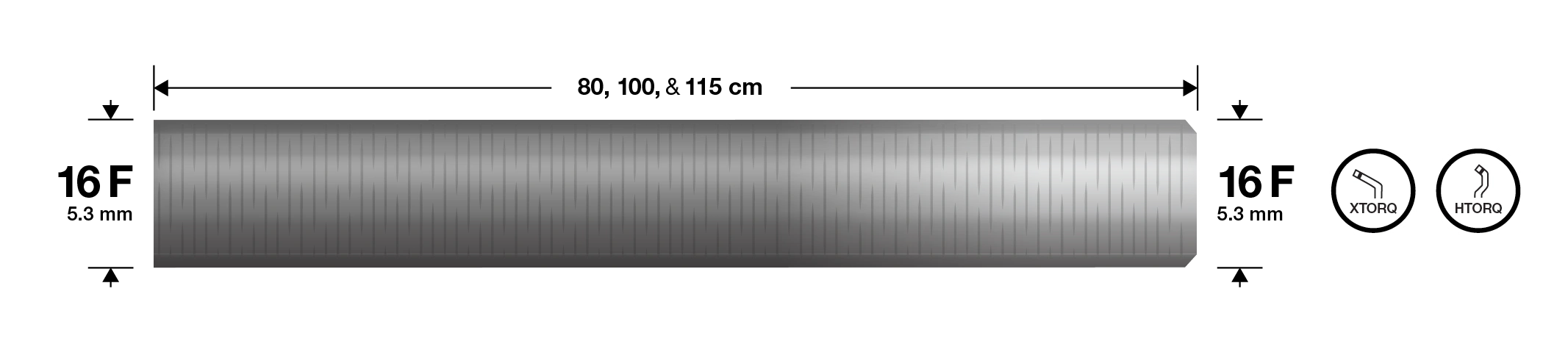
MaxID Technology
- Comparable to IDs of large-bore catheters while maintaining a lower profile
- Laser-cut stainless steel hypotube
a. Compared to Lightning Flash 2.0. Test performed and data on file at Penumbra Inc. Test performed using bovine blood and water. Bench test results may not be indicative of clinical performance.
- Compatible with Penumbra’s 17 F Element™ Vascular Access System
- 16 F sheath compatibility
- 80 cm (HTORQ Tip), 100 cm (XTORQ and HTORQ Tips), and 115 cm (HTORQ Tip) working lengths
Packaged with Select +™
Engineered for the pulmonary anatomy
- Available with H1 tip shape
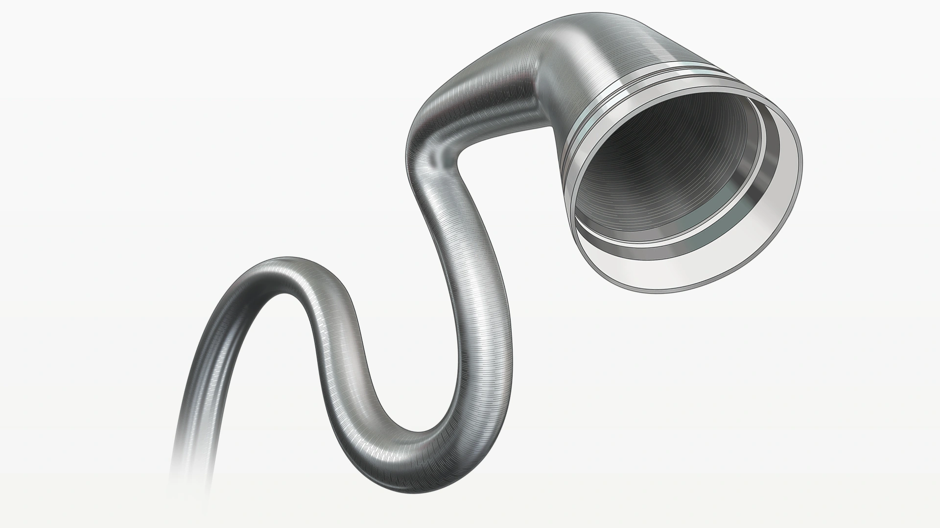
- Seamless Transition: 98% lumen occupancyb
- Designed for swift pulmonary selection

b. Data on file at Penumbra, Inc. Percentage reflects Lightning Flash Catheter lumen occlusion.

Lightning Flash 2.0: Next-Gen Software for the Treatment of PE
CIT, May 2024
Penumbra’s Dedication to Innovation: Expanding the CAVT™ Platform
EVT, Jan 2025

Product Gallery
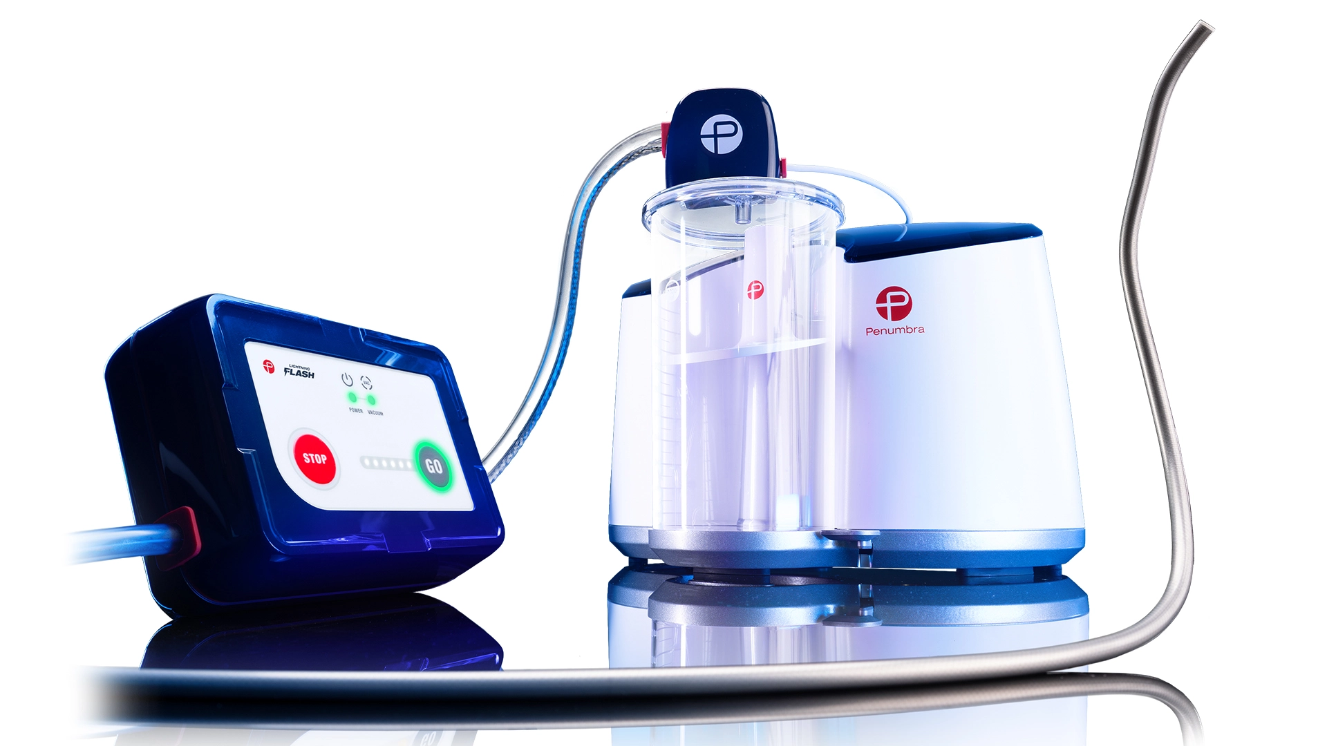
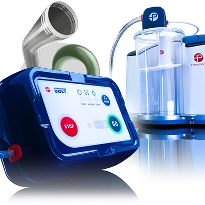
– Lightning Flash 3.0
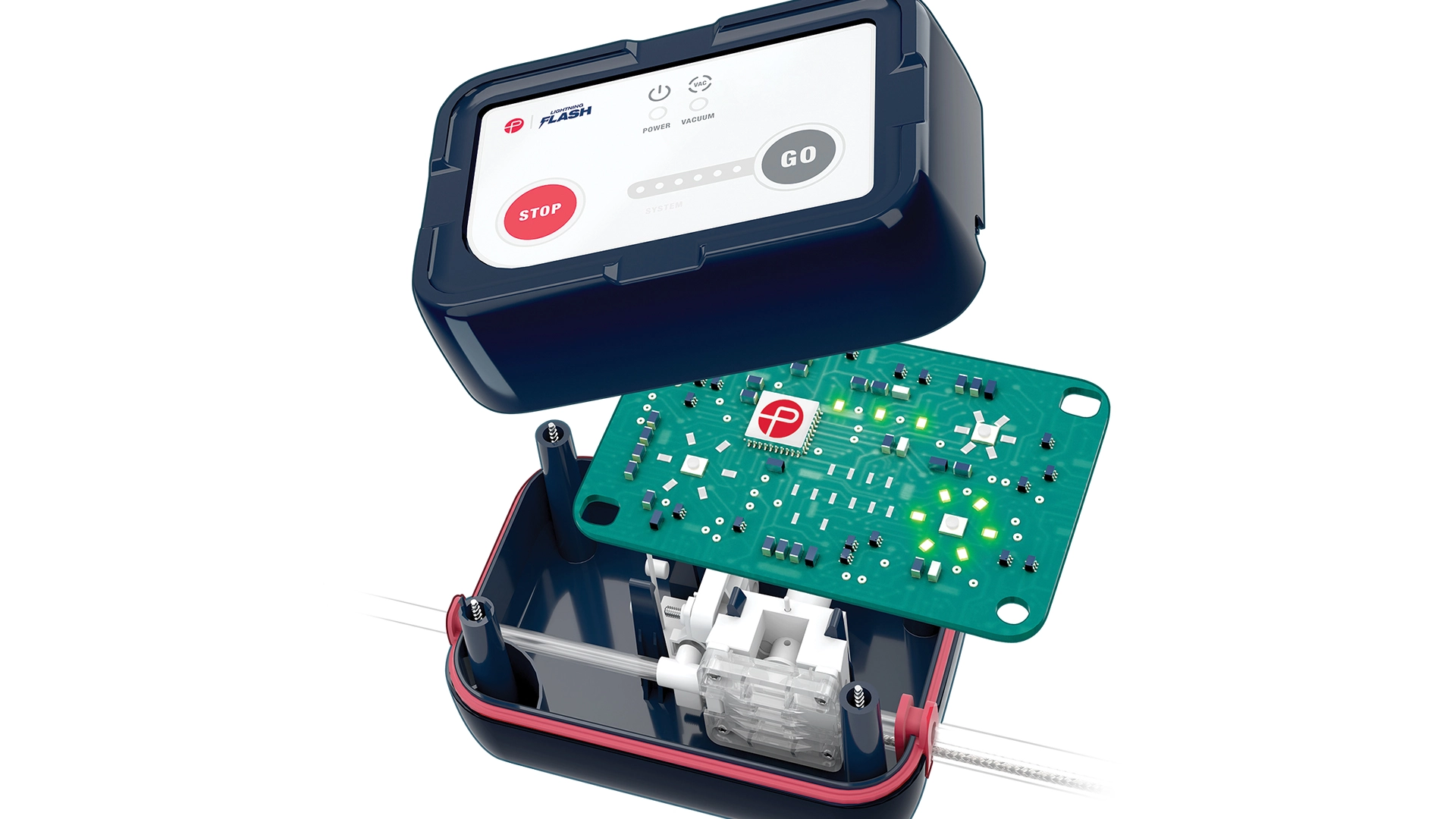
– Lightning Flash 3.0 Interface: Computer Assisted Vacuum Thrombectomy
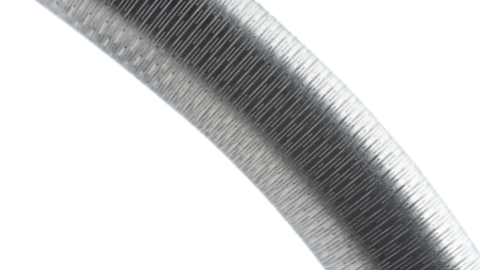
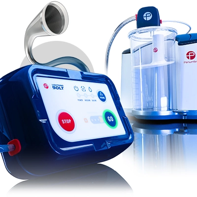
– Stainless Steel Hypotube
– CAT16 with Select +
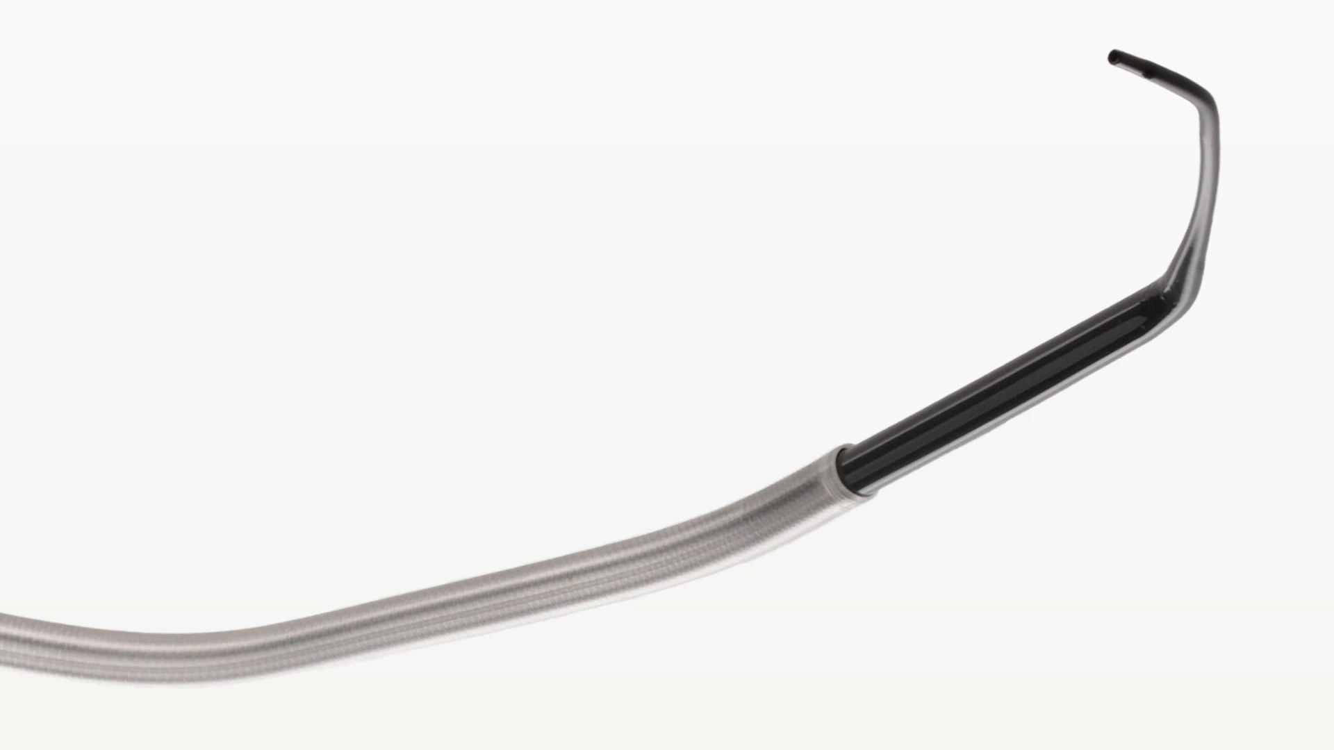
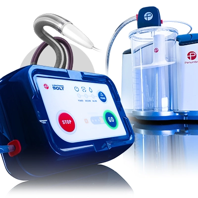
– Lightning Flash Aspiration Catheter
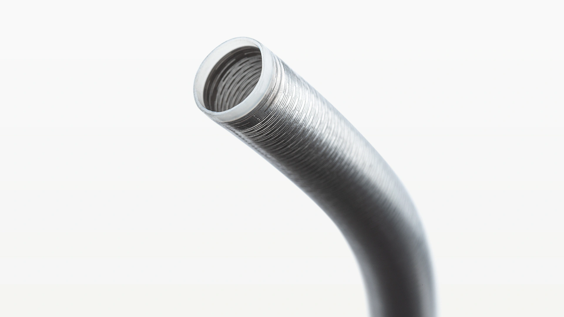
– Lightning Flash Aspiration Catheter Tip Shape
Please refer to Penumbra’s image license for use of materials
Pulmonary Embolism Cases
Lightning Flash® 3.0 – Bilateral Pulmonary Thrombectomy
Dr. Tony Lu
Houston Methodist
Houston, TX


Lightning Flash® 3.0 – Bilateral Pulmonary Embolism Thrombectomy
Dr. Matt Ramsey
Duke University Hospital
Durham, NC


Venous Cases
Lightning Flash® 3.0 – Venous Thrombectomy
Dr. Brad Grimsley
Baylor The Heart Hospital
Dallas, TX


Lightning Flash® 3.0 – Venous Thrombectomy
Dr. Brett Hyatt
Mississippi Baptist Hospital
Jackson, MS


Images used with permission and provided for illustrative purposes only. Procedural techniques and decisions based on physician’s medical judgment. Individual results may vary. Consents on file at Penumbra, Inc.

Related Articles

In the News
Read our latest news, articles, and posts.
Meetings & Events
View our conference and event schedule.
Our Impact
Learn how our latest innovations are transforming patient care.
Clinical Evidence
Access the latest clinical research and data.

Request a Demo
Want to see Penumbra products in action? Request a demonstration for your facility.