
Meeting the Rising Tide of Chronic Subdural Hematoma with Evolving Procedural Options
Chronic subdural hematoma (cSDH) is the most common neurosurgical condition affecting older adults — and its incidence continues to rise rapidly as the population ages.1 Researchers predict that within the next decade the growth in cSDH frequency will place increasing strain on neurosurgical services and healthcare systems worldwide.
As clinicians seek safe, long-lasting solutions for an often frail patient population, emerging minimally invasive approaches such as middle meningeal artery (MMA) embolization have shown promise, and multiple large, randomized trials are currently underway.2
What Is a Subdural Hematoma?
A subdural hematoma (SDH) occurs when blood accumulates between the dura mater — the protective layers that surround your brain — and the surface of the brain. This abnormal collection of blood damages cells in the brain lining, causing inflammation and excessive cell growth.
While acute SDH is commonly caused by traumatic injury, older adults experience much higher rates of chronic subdural hematomas than the general population.1,3 Age-related brain atrophy stretches bridging veins, making them more susceptible to rupture, even after minor or unnoticed trauma, leading to gradual symptom onset.4
SDH Signs and Symptoms
Symptoms may appear immediately, or develop gradually in the case of cSDH. Presentation varies depending on hematoma size and location but may include:4
- Confusion or coma
- Decreased memory
- Problem speaking or swallowing
- Balance or walking problems
- Drowsiness
- Headache
- Seizures
- Weakness or numbness of arms, legs or face
- Nausea/Vomiting
- Aphasia, a language disorder that impacts communication
- Visual difficulties
- Pupil size
- Ringing sound in the ears
The Growing Incidence of Chronic Subdural Hematoma
Chronic subdural hematoma is the most common neurosurgical diagnosis in seniors, with incidence increasing in parallel with life expectancy. Recurrence of the disease is common even with conventional management.2 Researchers estimate that by the end of the decade, cSDH will become the most common craniosurgical disease worldwide.2 In recent years, middle meningeal artery (MMA) embolization has shown promise in reducing recurrence and minimizing the need for repeat surgeries.2 Procedural volumes are projected to exceed 140,000 cases annually in older adults — potentially outpacing even large vessel stroke cases — underscoring the need for scalable, minimally invasive options.1
SDH Management Options
- MMA Embolization
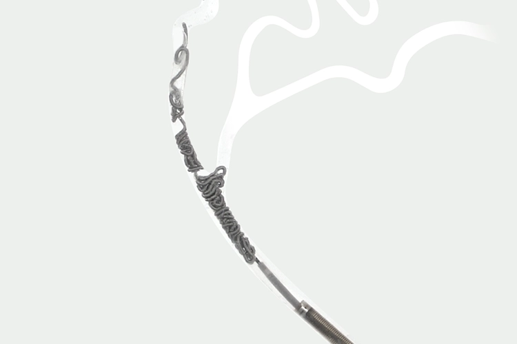
MMA embolization targets the vascular supply responsible for sustaining and recurring chronic subdural hematomas (cSDH). The MMA provides blood supply to the dura mater and feeds the membrane capillaries supplying the cSDH. This minimally invasive procedure blocks blood flow to this region, ultimately allowing the hematoma to be absorbed and decrease in size. The procedure is performed endovascularly, typically via femoral or radial artery access. Under fluoroscopic guidance, a microcatheter is navigated to the MMA, where embolic materials are deployed to close off the artery, helping in hematoma resolution.5Types of embolics include:
- Platinum coils
- Particulate embolics
- Liquid embolic agents
MMA embolization is increasingly used as a primary option, adjunct to surgery, or prophylactic approach following burr hole drainage.
- Craniotomy
This surgical procedure involves the removal of a section of the skull to access and extract the hematoma.5 The surgeon makes a cut through the scalp to expose the bone. A high-speed drill and saw may be used to cut a portion of the bone to access the brain, which is usually placed back in place at the end of the procedure.6
- Burr Hole Craniostomy
Burr hole drainage is the most commonly used surgical intervention for cSDH. During this surgical procedure, one or more small holes are drilled into the skull. A tube is then inserted through the hole to help remove the blood. A subdural drainage system is often employed during the postoperative period.5
Case Study: Successful MMA Embolization for Chronic Subdural Hematoma
Dr. Allison Strickland, MD, a dual-trained neurosurgeon at University of Mississippi Medical Center in Jackson, MS, shares a case of a bilateral middle meningeal artery (MMA) embolization of a 60-year-old male patient with chronic subdural hematoma.
Case Presentation
A 60-year-old male with HIV and poor functional baseline presented to the emergency department with new onset seizures. A CT of the head demonstrated large acute on chronic right-sided subdural hematoma and small chronic left-sided subdural hematoma. Patient was not a candidate for open surgery, and was taken for bilateral middle meningeal artery embolization with SwiftPAC™.
Intervention
Access was obtained using the BENCHMARK™ 071 access catheter. The MIDWAY™ 43 delivery catheter was advanced over a 10-system microcatheter and an 0.014” guidewire into the right MMA. Bilateral MMA embolization was performed by placing one 10 cm SwiftPAC Packing Coil and one 15 cm SwiftPAC Packing Coil in the right MMA. The procedure was repeated on the left side with one 20 cm SwiftPAC Packing Coil and one 1 mm x 5 cm SwiftSET™ Coil were placed in the left MMA.
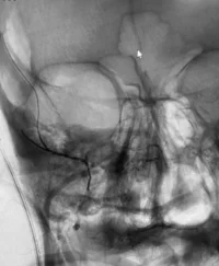
POST: Interval coil embolization of the right MMA with SwiftPAC Packing Coils
Discussion
The patient stopped seizing and was discharged home with family. The large acute on chronic right-sided subdural hematoma decreased in size and was smaller on follow-up imaging.
“In MMA embolization, we are working in a complex vascular network with critical anastomoses that require careful consideration. SwiftPAC coils provide a controlled approach to MMA embolization for targeted occlusion that minimizes risk to the patient,” said Dr. Strickland.
Learn more about the Swift Coil System.
Important Safety Information
Additional information about Penumbra’s products can be located on Penumbra’s website at https://www.penumbrainc.com/products/neuro-embolization-system/. Caution: Federal (USA) law restrictions these devices to sale by or on the order of a physician. Prior to use, please refer to Instructions for Use (IFU) for complete product indications, contraindications, warnings, precautions, potential adverse events, and detailed instructions for use. For the complete Penumbra IFU Summary Statements, visit: peninc.info/risk. Please contact your local Penumbra representative for more information.
Copyright ©2026 Penumbra, Inc. All rights reserved. SwiftPAC, BENCHMARK, MIDWAY, and SwiftSET are registered trademarks or trademarks of Penumbra, Inc. in the USA and other countries.
1. Rai AT, Halak AA, Lakhani DA, et al. Population-based estimates suggest middle meningeal artery embolization for subdural hematomas could significantly expand the scope of neurovascular therapies. J Neurointerv Surg. 2024 Apr 11:jnis-2024-021686. doi:10.1136/jnis-2024-021686.
2. Chen H, Colasurdo M, Malhotra A, et al. Advances in chronic subdural hematoma and membrane imaging. Front Neurol. 2024 Apr 25;15:1366238. doi:10.3389/fneur.2024.1366238.
3. Pierre L, Kondamudi NP. Subdural Hematoma. In: StatPearls. Treasure Island (FL): StatPearls Publishing; August 12, 2023.
4. Nouri A, Gondar R, Schaller K, Meling T. Chronic Subdural Hematoma (cSDH): A review of the current state of the art. Brain Spine. 2021;1:100300. Published 2021 Nov 2. doi:10.1016/j.bas.2021.100300.
5. Désir LL, D’Amico R, Link T, et al. Middle Meningeal Artery Embolization and the Treatment of a Chronic Subdural Hematoma. Cureus. 2021; 13(10):e18868. Published 2021 Oct 18. doi:10.7759/cureus.18868.
6. National Library of Medicine (US). Brain surgery. MedlinePlus Medical Encyclopedia. https://medlineplus.gov/ency/article/003018.htm. Accessed March 25, 2026.
Related Articles
-
Young, Healthy, and at Risk: These Patient Stories Show Blood Clots Don’t Discriminate
October 12, 2026 -
CIRSE 2026: New STRIKE-PE Analysis Shows Sustained Improvements in Functional Outcomes and Quality of Life One Year After CAVT Treatment
October 7, 2026 -
A Patient Was Told Her Pelvic Pain Was “Normal” for Years. It Wasn’t.
September 25, 2026 -
Employee Spotlight: Dillon Gedeon
September 23, 2026